


Steven Rogers (Manchester, UK) presented the case for using contrast-enhanced tomographic 3D ultrasound imaging for peripheral arterial disease, telling the audience at Charing Cross Symposium (CX; 15–18 April, London, UK): “Our surgeons certainly prefer the 3D vein mapping to a 2D vein map”.
According to Rogers, tomographic 3D ultrasound imaging is “minimally-invasive, you can do it with relatively little experience, it is fast, it is inexpensive, it can be reimbursed, and potentially it is becoming more clinically valuable; perhaps it is the way forward.”
The multi-census study, funded by a European Union grant, investigated the use of contrast-enhanced ultrasound imaging in below-the-knee vessels for patients with peripheral arterial disease. So far, Rogers and colleagues have recruited 100 patients (including 414 arteries). All patients underwent some form of angiogram, whether that be CT, MR or catheter, and then within the week also had a tomographic ultrasound scan “from popliteal to toes”. Rogers told delegates that they chose to image below-the-knee arteries as he says “standard duplex is good enough for the femoropopliteal segment”, and the investigators wanted to “address the areas where it is weak”, which Rogers claims is below the knee.
To take the scan, the physicians move the probe, which has sensors attached to it, down the leg, and take a two dimensional ultrasound image. Showing such a visualisation on the screen, Rogers said “I defy anybody to give me a diagnosis from this standard contrast 2D image”. Explaining the next steps, he continued: “We compute that [two dimensional image] into the third dimension; then we can rotate it and review it from any angle. We can also see collateral vessels that feed the muscles, and we can see potential areas of disease.”
To assess the reproducibility of the tomographic ultrasound scan, Rogers explained that they set out to determine the degree of intraoperative observer agreement when making diagnoses based off the images. He reported finding a high degree of intraoperative observer agreement, and added that as “the confidence intervals are relatively narrow, we know there is a degree of precision in its reproducibility”.
However, Rogers did acknowledge some limitations of this imaging modality. As this is an ultrasound technique, he admits that “we are still constrained by the sonic constraints of ultrasound”, and says for this reason “deep vessels in particular are challenging, as are calcified vessels”. Notwithstanding these restrictions to its use, Rogers believes the minimally-invasive, 3D mapping technique “may well be a go to test in the future arsenal for vascular physicians”, although “there is still a long way to go”.
Session chair Robert Morgan (London, UK) asked Rogers whether this imaging tool was an ultrasound machine “just for vascular imaging”, or if it is “a general ultrasound machine with tomographic capabilities?”
“It is a bolt on, basically. You need a normal duplex instrument with contrast options, and it works by screen capture, so you just plug in an HDMI cable. At the minute it is a separate system—the current system is £44,000, they have, literally about 10 minutes ago, launched the second generation device, which costs £9,000 and plugs into a standard laptop.”
After summarising “The point is to find if we have got a binary restenosis; a residual or a current stenosis that is haemodynamically significant”, moderator Andrew Holden (Auckland, New Zealand) questioned whether Rogers felt confident that, using this technique, the speaker felt able to “differentiate between a 30–40% residual as compared to a 70% residual with the imaging data”.
Rogers was clear: “We can certainly measure the percentage, whether it is 30, 40 or 70”; he added: “I think the key here is that we can use it to go back and re-target the duplex and take the velocity shift across a particular area of interest, so this imaging tool is complementary [to current imaging techniques] rather than offering a replacement right now”.
Source VascularNews
Duc Tin Clinic
Tin tức liên quan
Performance diagnostique de l’interféron gamma dans l’identification de l’origine tuberculeuse des pleurésies exsudatives
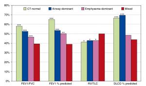
A Mixed Phenotype of Airway Wall Thickening and Emphysema Is Associated with Dyspnea and Hospitalization for Chronic Obstructive Pulmonary Disease.
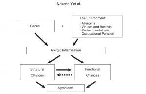
Radiological Approach to Asthma and COPD-The Role of Computed Tomography.
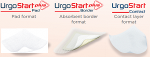
Significant annual cost savings found with UrgoStart in UK and Germany
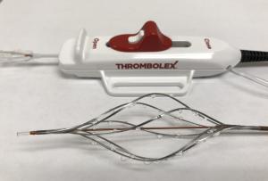
Thrombolex announces 510(k) clearance of Bashir catheter systems for thromboembolic disorders
Phone: (028) 3981 2678
Mobile: 0903 839 878 - 0909 384 389