Vascular remodelling may occur in response to sustained, regular exercise, suggests a study of aortic dilatation among long-term endurance athletes. Timothy W Churchill (Massachusetts General Hospital, Boston, USA) et al found a “marked increase in the prevalence of aortic dilatation based on established population nomograms. This finding was consistent between men and women, and across athletes participating in two of the most common endurance sports [running and rowing]”. The elevated prevalence was found without clear explanatory risk factors.
Writing in JAMA Cardiology, Churchill and colleagues say: “It appears that the aorta is an exercise-responsive plastic organ that remodels in the setting of long-term exercise. Further longitudinal study will be required to establish definitive clinical correlates of these findings.”
The study aimed to assess the prevalence of aortic dilatation among long-term masters-level male and female athletes with about two decades of exercise exposure. Before the present cross-sectional study, the distribution of aortic sizes in veteran endurance athletes was unknown, as was whether it represents aortic adaptation to long-term exercise, similar to that of ventricular remodelling.
Researchers evaluated aortic size in masters-level rowers and runners aged 50–75 years enrolled from competitive athletic events across the USA between February and October 2018. The primary outcome was aortic size at the sinuses of Valsalva and the ascending aorta, measured using transthoracic echocardiography. Aortic dimensions were compared with age, sex, and body size-adjusted predictions from published nomograms, and z scores were calculated where applicable.
Among 442 athletes (mean age 61 years, standard deviation [SD] 6; 267 men [60%]; 228 rowers [52%], 214 runners [48%]), clinically relevant aortic dilatation, defined by a diameter at sinuses of Valsalva or ascending aorta of ≥40mm, was found in 21% (n=94) of all participants (83 men [31%] and 11 women [6%]). The authors say it represents “a marked increase in the prevalence of aortic dilatation compared with that predicted by established age- and sex-specific population nomograms [all p<0.001]”. Overall, 105 individuals (24%) had at least one z score of 2 or more, indicating an aortic measurement greater than two SDs above the population mean. In multivariate models adjusting for age, sex, body size, hypertension, and statin use, both elite competitor status (rowing participation in world championships or Olympics or marathon time under two hours and 45 minutes) and sport type (rowing) were independently associated with aortic size.
The study team say that the findings have “potentially important clinical implications”, and “fill an important gap in our understanding of how long-term participation in endurance sport affects the cardiovascular system”.
They speculate that mild to moderate dilatation of the ascending aorta among long-term exercisers may “represent a previously unrecognised and benign adaptation to sport, similar to exercise-induced eccentric left ventricular (LV) hypertrophy”.
An alternative hypothesis is that “aortic dilatation in the setting of long-term endurance training may represent a novel form of acquired overuse pathology with attendant implications on morbidity and mortality”.
Because the study excluded athletes with bicuspid aortic valves, to maximally isolate the effect of long-term exercise, the authors suggest that the findings may be “conservative estimates for the overall population of masters-level athletes”.
But, as experienced well-trained athletes participating in two common endurance sports were assessed, Churchill et al caution that the findings may not be generalisable to more recreational exercisers, and to athletes participating in other endurance disciplines. Other limitations are that the study was cross-sectional and thus “can neither establish causality between exercise and aortic dilatation nor permit conclusions about the natural history and clinical risk profile of aortic dilatation among long-term competitive athletes”.
In addition, exercise exposure measures did not include data defining exercise training intensity and blood pressure response to exercise, “both of which may represent mechanistic underpinnings of our findings”, say the researchers. And, because those taking echocardiographic measurements were not blinded to the sport status of participants, bias may have been introduced.
“Future studies aimed at defining the natural history of aortic dilatation in this population with an emphasis on clinical outcomes, including the incidence of acute aortic syndromes and elective aortic surgical intervention, will be required to resolve this fundamental uncertainty,” conclude the investigators. “In the absence of such data, clinical implications of our findings remain uncertain and will require individualised assessment.”
Source CardiovascularNews
Duc Tin Clinic
Tin tức liên quan
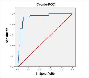
Performance diagnostique de l’interféron gamma dans l’identification de l’origine tuberculeuse des pleurésies exsudatives
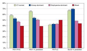
A Mixed Phenotype of Airway Wall Thickening and Emphysema Is Associated with Dyspnea and Hospitalization for Chronic Obstructive Pulmonary Disease.

Radiological Approach to Asthma and COPD-The Role of Computed Tomography.

Significant annual cost savings found with UrgoStart in UK and Germany
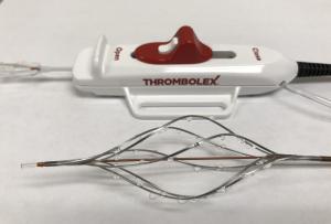
Thrombolex announces 510(k) clearance of Bashir catheter systems for thromboembolic disorders
Phone: (028) 3981 2678
Mobile: 0903 839 878 - 0909 384 389
