


Identification and treatment of vascular malformations is a challenging endeavor for physicians, especially given the great concern and anxiety created for patients and their families. The goal of this article is to provide a review of vascular malformations, organized by subtype, including capillary, venous, lymphatic and arteriovenous malformations. Only by developing a clear understanding of the clinical aspects, diagnostic tools, imaging modalities, and options for intervention will appropriate care be provided and results maximized.
Vascular anomalies, a group encompassing a wide variety of lesions related to the disorder of vascular development, remain both diagnostic and treatment challenges to treating physicians. The terminology used to describe and classify vascular anomalies is the key for proper diagnosis and treatment. The classification system established by the International Society for the Study of Vascular Anomalies (ISSVA) is now a widely accepted system used to categorize vascular anomalies into two types: (1) vasoproliferative or vascular neoplasms such as hemangioma, and (2) vascular malformations.1
The distinction between the two is based on histopathological assessment of increased cell turnover. Vascular tumors, formerly classified as hemangiomas, are true neoplasms with pathologic cell proliferation. These tumors typically exhibit rapid postnatal growth and slow regression into late childhood.2 Vascular malformations, on the other hand, are comprised of abnormally formed channels within a vascular apparatus that are lined by endothelial cells and do not undergo abnormal cellular turnover. They too are congenital in nature, but often go unnoticed at birth, never regress, and grow proportionally with the individual.3
In this article, we will focus solely on vascular malformations, reviewing the basic nomenclature, etiology, and diagnostic criteria for each subcategory and discussing treatment options available today. An in-depth review of vascular malformations associated with known syndromes as well as vascular tumors will be provided in separate articles. It is important to be able to make accurate diagnosis, understand the basic physiology, and use appropriate diagnostic and treatment modalities to optimize outcome.
Vascular Malformations
Vascular malformations are grouped together based upon their common embryological origin of having a single endothelial cell lining.3 Vascular malformations are thought to result from developmental errors during embryogenesis, such as abnormal signaling processes that control apoptosis, maturation, and growth of vascular cells. These errors lead to the persistence of vascular plexus cells with a certain degree of differentiation.4 There are four major categories of vascular malformations based on their flow characteristics: slow-flow (capillary malformation, venous malformation, lymphatic malformation) and fast-flow (arteriovenous malformation). These lesions often have components of multiple malformations, such as a mixed lymphatico-venous malformation, further adding to the confusion with respect to proper nomenclature
As with any medical or surgical anomaly, accurate diagnosis is paramount to successful treatment. With vascular malformations, a thorough history and physical exam will allow the astute physician to make a sound diagnosis for a majority of the clinical cases presented. Additionally, proper imaging modalities such as ultrasound (US) with gray scale, color Doppler and spectral Doppler tracings, or magnetic resonance imaging (MRI) can aid when the diagnosis is in question, as there may be overlap in clinical appearance in these anomalies.5 These imaging modalities can assist in confirming particular attributes of the lesion, defining anatomic locations/boundaries, and planning potential surgical intervention.
In general, the management of vascular malformation is expectant in nature with both noninvasive and invasive treatment of symptomatic lesions. Lesions located in the head and neck region, however, require special attention as they can cause obstruction of critical structures such as the visual axis or the airway

Very large lymphatic malformation involving the right head and neck region and compressing the airway.
Capillary Malformation
Capillary malformations (CMs) affect the capillaries in the papillary dermis and commonly appear as a macular, pink or purple stain that is present at birth and persists throughout life.6 Capillary malformations, found in 0.5% of the population, were initially referred to as “port-wine stain,” which is inaccurate, but has persisted due to its widespread use throughout the literature. The majority of CMs appear in the face and tend to be in the trigeminal nerve distribution, especially ophthalmic (V1) and maxillary (V2) divisions.7Capillary malformations in the V1 or midline distribution should alert physicians of possible central nervous system (CNS) involvement and warrant imaging studies as this is highly associated with leptomeningeal involvement and subsequent seizure disorders (i.e,, Sturge-Weber syndrome).
As the patient progresses into adulthood, the stains tend to darken and thicken into a “cobblestone” appearance and can distort facial features, including the underlying bony structures. There is currently no imaging modality needed to assist in diagnosis, but MRI is strongly recommended to rule out CNS involvement. Positive findings include gyral enhancement, enlargement and enhancement of ipsilateral choroid plexus, progressive cortical atrophy and calcification.8 Cerebral angiography can detect parenchymal contrast stasis and abnormal cortical veins associated with CNS involvement.
The majority of treatment modalities are ablative in nature, such as the pulsed-dye laser (580–595 nm).9 Significant improvement in the color of the stain can be seen when laser treatment is begun in infancy and when applied to CMs of the lateral face. Noticeable lightening is typically seen in greater than 75% of patients. Better results are achieved when therapy is initiated early, thus it is recommended that treatment commence before 6 months of age. Surgery is reserved for lesions that are refractory to ablative treatment or are causing significant disfigurement (Fig. 3).10

Tin tức liên quan
Performance diagnostique de l’interféron gamma dans l’identification de l’origine tuberculeuse des pleurésies exsudatives
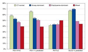
A Mixed Phenotype of Airway Wall Thickening and Emphysema Is Associated with Dyspnea and Hospitalization for Chronic Obstructive Pulmonary Disease.

Radiological Approach to Asthma and COPD-The Role of Computed Tomography.
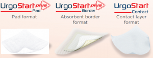
Significant annual cost savings found with UrgoStart in UK and Germany
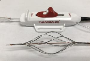
Thrombolex announces 510(k) clearance of Bashir catheter systems for thromboembolic disorders
Phone: (028) 3981 2678
Mobile: 0903 839 878 - 0909 384 389