During the prize session at this year’s European Society for Vascular Surgery annual meeting (ESVS Month, 29 September–29 October, virtual), Maaz Syed (University of Edinburgh, Edinburgh, UK) presented early clinical outcomes of a “promising multimodality imaging technique” in acute aortic syndrome—18F-sodium fluoride positron emission tomography (PET).
Despite intensive imaging surveillance and aggressive therapy, mortality from acute aortic syndrome has remained static, Syed relayed, emphasising a “pressing need” to identify those patients most at risk of the “rare but catastrophic” conditions that are aortic dissection, intramural haematoma, and penetrating ulcer.
Syed and the University of Edinburgh research team believe that combining the power of computed tomography (CT) with information on the biological processes that drive morphological change “holds great promise” in this field.
In their study, supported by the British Heart Foundation, Syed et al investigate microscopic calcification. Explaining the rationale behind this, he detailed that microcalcification is “beyond the resolution of conventional CT, but can be detected using sodium fluoride, which binds to it”.
Syed informed viewers that using the radio isotope version of fluorine, 18F, the team can study the pattern of radioactive emission within the tissue to detect microcalcification. “This is the basis of 18F-sodium fluoride PET,” he clarified.
For this study, the research team invited all patients in Scotland over the age of 25 years with acute aortic syndrome for an 18F-sodium fluoride PET/CT, while healthy controls with normal sized aortas were recruited from the National Screening Programme. Syed and colleagues compared fluoride binding between groups.
Syed detailed that patients and healthy controls were matched for age, sex, BMI, and—on the whole—comorbidities and medications. The acute aortic syndrome group were more likely to smoke and have hypertension, he informed the audience.
On systematic assessment, the team found that patients with acute aortic syndrome had significantly greater fluoride binding than healthy controls, and amongst the three sub-pathologies it was the penetrating aortic ulcers that had the most intense fluoride uptake, followed by aortic dissections and then intramural haematomas.
Syed noted an “interesting finding” was that when compared to proximal healthy aorta, sodium fluoride binding concentrated at the site of intimal disruption.
“We observed no significant difference in fluoride binding between patients in the subacute and chronic groups, and fluoride binding was also similar across Stanford classification,” he added.
Moving on to results, Syed stressed that while the research group have less than one-year follow-up results at present, this can still provide an “indicator” of emerging clinical trends.
The team devised a simple linear regression model to study the interaction between false lumen fluoride binding and clinical events, which showed that fluoride binding intensity became a “significant predictor” of clinical events after adjusting for patient sex. In addition, the relationship persisted independent of patients’ age and aortic diameter, Syed reported.
Looking to the future, Syed posited: “If we use this information to predict fluoride binding in the subacute phase of an aortic dissection, we can be fairly confident that fluoride binding would be greater in those that experienced aortic rupture or repair versus those that remained event free.”
Summarising the key findings of the early clinical outcomes of this study, Syed relayed three main takeaways: first, 18F-sodium fluoride binds preferentially to the aortas of patients with acute aortic syndrome; second, the distribution of 18F-sodium fluoride suggests peak binding activity is at the site of intimal disruption; and, finally, preliminary findings from early follow-up suggest that acute dissection patients with high false lumen fluoride uptake may also develop clinical events.
“This is the first characterisation of sodium fluoride PET/CT in patients with acute aortic syndrome,” Syed claimed, describing it as a “promising multimodality imaging technique that detects aortic cellular injury and predicts clinical events”.
Closing his presenting, Syed was confident about future applications of this technology: “We believe that combining established clinical and morphological predictors with sodium fluoride PET may one day help us better identify the vulnerable aorta in patients with acute aortic syndrome. Much work is still required, but we can see this potential.”
Source VascularNews
Duc Tin Clinic
Tin tức liên quan
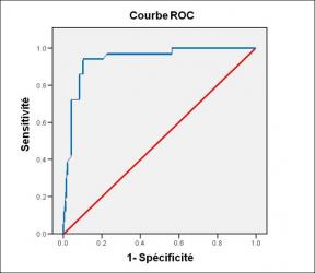
Performance diagnostique de l’interféron gamma dans l’identification de l’origine tuberculeuse des pleurésies exsudatives
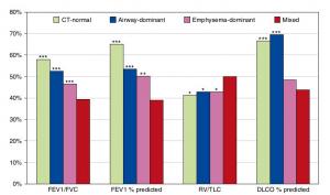
A Mixed Phenotype of Airway Wall Thickening and Emphysema Is Associated with Dyspnea and Hospitalization for Chronic Obstructive Pulmonary Disease.

Radiological Approach to Asthma and COPD-The Role of Computed Tomography.
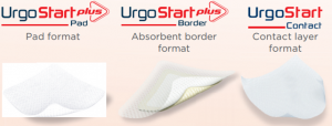
Significant annual cost savings found with UrgoStart in UK and Germany
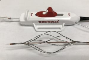
Thrombolex announces 510(k) clearance of Bashir catheter systems for thromboembolic disorders
Phone: (028) 3981 2678
Mobile: 0903 839 878 - 0909 384 389
