Lab-created heart valves implanted in young lambs were capable of growth within the recipient, a study published in Science Translational Medicine has found. The valves also showed reduced calcification and improved blood flow function compared to animal-derived valves currently used when tested in the same growing lamb model, researchers at the University Of Minnesota Twin Cities, Minneapolis, USA, who conducted the study, reported.
If confirmed in humans, lab-grown valves could prevent the need for repeated valve replacement surgeries, for example in children with congenital heart defects, the study’s authors suggest. Additionally they report that the valves can be stored for at least six months, which means they could provide surgeons with an “off the shelf” option for treatment.
“This is a huge step forward in paediatric heart research,” said Robert Tranquillo, the senior researcher on the study and a University of Minnesota professor in the Departments of Biomedical Engineering and the Department of Chemical Engineering and Materials Science. “This is the first demonstration that a valve implanted into a large animal model, in our case a lamb, can grow with the animal into adulthood. We have a way to go yet, but this puts us much farther down the path to future clinical trials in children. We are excited and optimistic about the possibility of this actually becoming a reality in years to come.”
Currently, researchers have not been able to develop a heart valve that can grow and maintain function for paediatric patients. The only accepted options for these children with heart defects are valves made from chemically treated animal tissues that often become dysfunctional due to calcification and require replacement because they do not grow with the child. These children will often need multiple surgeries until a mechanical valve is implanted in adulthood. This requires them to take blood thinners the rest of their lives.
In this study, Tranquillo and colleagues used a hybrid of tissue engineering and regenerative medicine to create the growing heart valves. Over an eight-week period, they used a specialised tissue engineering technique they previously developed to generate vessel-like tubes in the lab from a post-natal donor’s skin cells. To develop the tubes, researchers combined the donor sheep skin cells in a gelatin-like material, called fibrin, in the form of a tube and then provided nutrients necessary for cell growth using a bioreactor.
The researchers then used special detergents to wash away all the sheep cells from the tissue-like tubes, leaving behind a cell-free collagenous matrix that does not cause immune reaction when implanted. This means the tubes can be stored and implanted without requiring customised growth using the recipient’s cells, the researchers claim.
The next step was to precisely sew three of these tubes—about 16mm in diameter—together into a closed ring. The researchers then trimmed them slightly to create leaflets to replicate a structure similar to a heart valve about 19mm in diameter.
Source CardiovascularNews
Duc Tin Clinic
Tin tức liên quan
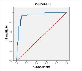
Performance diagnostique de l’interféron gamma dans l’identification de l’origine tuberculeuse des pleurésies exsudatives
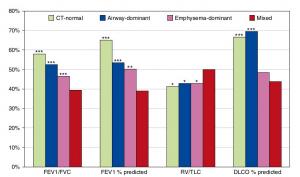
A Mixed Phenotype of Airway Wall Thickening and Emphysema Is Associated with Dyspnea and Hospitalization for Chronic Obstructive Pulmonary Disease.

Radiological Approach to Asthma and COPD-The Role of Computed Tomography.
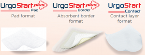
Significant annual cost savings found with UrgoStart in UK and Germany
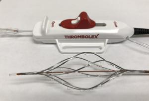
Thrombolex announces 510(k) clearance of Bashir catheter systems for thromboembolic disorders
Phone: (028) 3981 2678
Mobile: 0903 839 878 - 0909 384 389
