While there is large variability based on the site of insertion, patient characteristics and previous accesses, fluoroscopically-guided insertion of tunnelled central venous catheters (td-CVC) for dialysis can be considered a “low exposure” procedure, according to a report.
The report, which is published in The Journal of Vascular Access (JVA), also concludes, however, that patient exposure to ionising radiation during these dialysis access procedures is “significantly higher” than the levels associated with oncological port-a-cath CVC procedures—and nephrologists should be aware of the administered dose to ensure they comply with the 2013 European Directive on protection against ionising radiation exposure (IRE).
In concluding the report, Andreana De Mauri, the lead author and a researcher in the Nephrology and Dialysis Department at the Maggiore della Carità University Hospital (Novara, Italy), states: “In the next years, nephrologists have to take into account the legal implications, following the directives either of the scientific societies or governments, with regard to the high levels of competences, the clear definition of responsibilities and tasks of all professionals involved in the medical exposure, to ensure adequate protection of patients undergoing medical radio-diagnostic procedures.”
In spite of the indications laid out in the aforementioned 2013 European legislation, existing literature lacks information about the doses associated with fluoroscopically-inserted dialysis td-CVC, according to the report.
And, while td-CVC procedures are used worldwide—either for rescue vascular accesses or a first access in elderly and ill populations—the IRE related to them remains a knowledge gap among nephrologists, and clinical practitioners in general.
The researchers therefore conducted a retrospective study to quantify both the effective dose and the organ dose to relevant organs—including bone marrow, heart, lung, breast, thyroid and skin—in td-CVC, revising these procedures while taking into account radiation risk via dose-per-area product, fluoroscopy timings, and the different anatomical sites of catheter introduction.
The study involved 88 consecutive td-CVCs (Arrow Cannon II Plus, Teleflex), which were placed in 42 male patients (48%) and 46 female patients (52%). The anatomical districts they were used to access included a mix of the right internal jugular vein (RIJV), left internal jugular vein (LIJV), subclavian veins (SVs) and femoral veins (FVs).
Alongside this, the study also retrospectively revised 46 oncological port-a-cath CVC procedures acquired on Philips Healthcare’s Integris 5000 angiographic system, in order to compare the IRE levels in these procedures to those associated with td-CVC.
While the subsequent report concludes that fluoroscopically-guided td-CVC insertion procedures can be considered generally “low exposure”, with a minimal-to-very low risk of fatal cancer induction, it states that large variability and some exceptions exist in this area—mainly driven by the site at which the insertion took place, the characteristics of the patient, and the number of previous accesses carried out.
In addition, it states that the radiological exposure was similar for the LIJV, SV and FV access sites, but all of these were higher than the exposure in the RIJV and, as a result, the latter area should be preferred when the clinical conditions allow it.
The report also claims that, in the select few exceptional cases observed, organ doses are “not negligible” for procedures concerning the heart, lung, breast and bone marrow, and, as such, they should be added to the cumulative dose from all diagnostic and interventional procedures a dialysed patient is undergoing.
Due to the “minimal radiological risk” connected to td-CVC procedures, however, the report states that nephrologists should consider the “definitely superior benefit” deriving from this form of catheter insertion for patient dialysis—especially in patients without arteriovenous fistulae or a prosthetic loop on native vessels who require haemodialysis, as CVCs are mandatory here.
In spite of these minimal IRE-related risks, the report also concludes that these procedures should still be optimised where possible, as the nephrologist or other medical practitioner involved is still required to take part in this process by the 2013 European Directive. For example, it suggests that, depending on single-centre protocols, physicians could choose to insert a CVC using only ultrasound guidance and then perform the necessary radiological checks later via traditional chest radiography, for which the associated IRE is very low, and the related risks are negligible.
The report’s conclusion recommends that the nephrologist or practitioner in question should be aware of the ionising dose administered during a td-CVC procedure, so they can correctly inform the patient and ensure these details are included as part of the final report, as required by the law.
Source VascularNews
Duc Tin Clinic
Tin tức liên quan
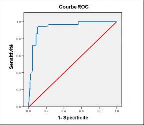
Performance diagnostique de l’interféron gamma dans l’identification de l’origine tuberculeuse des pleurésies exsudatives
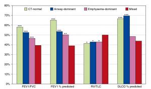
A Mixed Phenotype of Airway Wall Thickening and Emphysema Is Associated with Dyspnea and Hospitalization for Chronic Obstructive Pulmonary Disease.

Radiological Approach to Asthma and COPD-The Role of Computed Tomography.
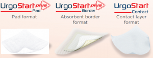
Significant annual cost savings found with UrgoStart in UK and Germany
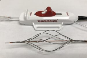
Thrombolex announces 510(k) clearance of Bashir catheter systems for thromboembolic disorders
Phone: (028) 3981 2678
Mobile: 0903 839 878 - 0909 384 389
