Four-dimensional (4D) flow magnetic resonance imaging (MRI) has the potential to identify patients with a higher risk of severe complications from aortic degeneration according to research published in JACC: Cardiovascular Imaging. The study employed a 4D flow MRI heatmap concept to detect abnormal aortic wall shear stress, a known stimulus for arterial wall dysfunction.
Aorta wall shear stress is the force exerted by blood flow on the arterial wall. It has been known that aortic 4D flow MRI can quantify regions exposed to high wall shear stress. Researchers at the Northwestern Medicine Department of Radiology (Chicago, USA), Bluhm Cardiovascular Institute (Chicago, USA) and the Libin Cardiovascular Institute (Calgary, Canada), Cumming School of Medicine (Calgary, Canada), University of Calgary (Calgary, Canada) evaluated the role of wall shear stress as a predictor of progressive aortic dilation in patients with bicuspid aortic valve disease. Progressive aortic dilation is associated with severe complications such as ascending aorta aneurysm, dissection, and rupture.
“Our findings indicate a potential role of 4D flow MRI derived wall shear stress as a new biomarker for arterial wall remodelling leading to higher rates of progressive aortic dilation in bicuspid aortic valve disease, thus exposing patients to a greater risk for aortic complications,” said Michael Markl, professor & vice chair for research, Departments of Radiology & Biomedical Engineering at Northwestern University. “This publication, based on innovative new 4D flow imaging developed at Northwestern Medicine, highlights the importance of PhD and MD cross-disciplinary collaboration that was instrumental for the success of this study.”
The study identified 72 bicuspid aortic valve patients who underwent MRI for surveillance of aortic dilation at baseline and follow-up five or more years later. Two patient groups were defined as slower or faster ascending aortic growth rates based on the mean growth rate of the cohort. For patients with higher rates of aortic dilation, 19.9 percent had elevated wall shear stress at baseline compared to 5.7 percent for those with slower growth rates.
“I think this a landmark paper for our group. It proves our concept that wall shear stress has a downstream effect on aortic degeneration. We are a step closer to using 4D MRI for clinical decision making,” said S Christopher Malaisrie, cardiothoracic surgeon, Northwestern Medicine Bluhm Cardiovascular Institute. The study was funded in part by the National Institutes of Health, and a fellowship by the the French College of Radiology Teachers and French Radiology Society.
“This publication showcases some very convincing data that is a capstone to our mission to validate 4D flow imaging as a clinical predictive tool for bicuspid aortopathy,” said Paul Fedak, director, Libin Cardiovascular Institute. “Clinicians can use this imaging tool and biomarker to help be more precise about prophylactic aortic resection.”
Source CardiovascularNews
Duc Tin Clinic
Tin tức liên quan
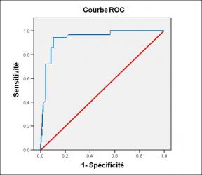
Performance diagnostique de l’interféron gamma dans l’identification de l’origine tuberculeuse des pleurésies exsudatives
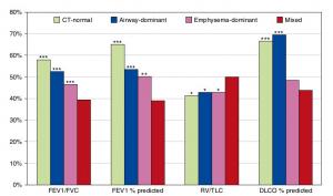
A Mixed Phenotype of Airway Wall Thickening and Emphysema Is Associated with Dyspnea and Hospitalization for Chronic Obstructive Pulmonary Disease.

Radiological Approach to Asthma and COPD-The Role of Computed Tomography.
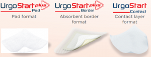
Significant annual cost savings found with UrgoStart in UK and Germany
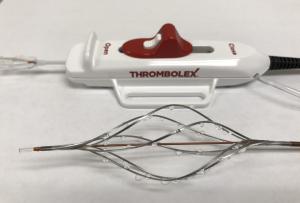
Thrombolex announces 510(k) clearance of Bashir catheter systems for thromboembolic disorders
Phone: (028) 3981 2678
Mobile: 0903 839 878 - 0909 384 389
