Siemens Healthineers has announced the launch of Artis icono biplane—an angiography system with detectors optimised in size for integrated use in the cath lab.
The system offers new features for diagnosing and treating cardiac arrhythmia, coronary heart disease, and structural heart disease that simplify clinical workflows and provides excellent image quality at a low radiation dose, Siemens said in a press release.
For complex cardiovascular diseases and their interventional treatment, Artis icono biplane allows for simple positioning of the C-arm—particularly for displaying images at steep angulations. In addition, images can be acquired simultaneously from different angles. This helps saving time and for patients has the potential to result in a lower contrast agent dose, Siemens adds in its release.
Artis icono biplane provides a type of X-ray exposure regulation that considers contrast-to-noise ratios to maintain the same high level of image quality, regardless of patient size and angulation. According to the company, the result is excellent image quality at a low radiation dose for physicians and patients.
Treatment of patients with cardiac arrhythmia requires electromapping of the heart to help the electrophysiologist identify the source of the arrhythmia and treat it through an ablation. Artis icono biplane will be compatible with mapping systems of other manufacturers, resulting in artifact-free images and the possibility to further reduce radiation exposure. Mapping systems typically use an electromagnetic field used for navigation, which usually leads to image artifacts on the angiography image. Instead of a clear map of the heart, images are then overlaid with stripes.
Artis icono biplane will provide system measures that may be able to reduce these image artifacts and allow for good visualisation of the devices being used, such as diagnostic catheters or ablation catheters.
In the case of coronary heart diseases, an integrated quantification feature of Artis icono biplane does away with the need for a preliminary examination with a pressure wire to determine stenosis relevance and subsequently, if a stent has to be placed.
A new feature “angio-derived vFFR”2 (Fractional Flow Reserve), means two images are needed to provide three-dimensional visualisation of the affected vessel and to obtain the necessary information.
According to Siemens, this image-guided method offers a couple of advantages: Even though using a pressure wire for diagnosis is a routine intervention, patients are still at risk for vascular injury. Furthermore, the administration of medications needed for the procedure can make patients feel unwell and also requires a certain amount of time to induce cardiac stress which is needed for the pressure wire examination.
![]() For interventions in patients with structural heart disease, such as transcatheter aortic valve implantation (TAVI), closure of the left atrial appendage (LAA closure), or transcatheter mitral valve implantation (TMVi), Artis icono biplane provides the option of performing fusion imaging. Angiography images can be overlaid with live ultrasound images These fused images help to ensure effective communication among treating interventional cardiologists and ecocardiographer.
For interventions in patients with structural heart disease, such as transcatheter aortic valve implantation (TAVI), closure of the left atrial appendage (LAA closure), or transcatheter mitral valve implantation (TMVi), Artis icono biplane provides the option of performing fusion imaging. Angiography images can be overlaid with live ultrasound images These fused images help to ensure effective communication among treating interventional cardiologists and ecocardiographer.
“Lack of time and cost pressure also affect the field of cardiology. With Artis icono biplane we are taking these two ongoing trends into account and are providing our customers with a feature-rich system for performing minimally invasive interventions that facilitates individual workflows and improves image quality in accordance with the ALARA [as low as reasonably achievable] principle,” says Doris Pommi, head of Cardiovascular Solutions at Siemens Healthineers.
Artis icono biplane can be adjusted to the individual user. As such, the user’s own established workflows can be defined, standardised, and stored to save valuable preparation time. If complications occur during vascular access, for example, one single click allows to select from as many as nine different automated system settings.
Since February 2022, the new system has been in pilot operation at the University Hospital of Innsbruck, Innsbruck, Austria.
Source CardiovascularNews
DUc TIn CLinic
Tin tức liên quan
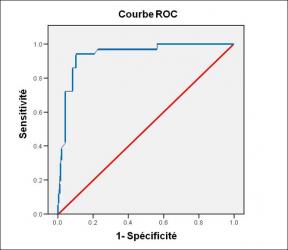
Performance diagnostique de l’interféron gamma dans l’identification de l’origine tuberculeuse des pleurésies exsudatives
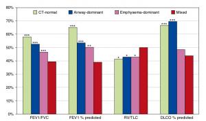
A Mixed Phenotype of Airway Wall Thickening and Emphysema Is Associated with Dyspnea and Hospitalization for Chronic Obstructive Pulmonary Disease.

Radiological Approach to Asthma and COPD-The Role of Computed Tomography.
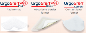
Significant annual cost savings found with UrgoStart in UK and Germany
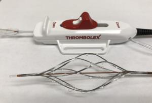
Thrombolex announces 510(k) clearance of Bashir catheter systems for thromboembolic disorders
Phone: (028) 3981 2678
Mobile: 0903 839 878 - 0909 384 389
