The diagnosis of Hypertrophic Cardiomyophathy (HCM) rests on the detection of increased Left Ventricular (LV) wall thickness by any imaging modality.
The diagnosis of Hypertrophic Cardiomyophathy (HCM) rests on the detection of increased Left Ventricular (LV) wall thickness by any imaging modality.
- In adults, HCM is defined by a wall thickness ≥15 mm in one or more LV myocardial segments—as measured by any imaging technique (echocardiography, cardiac magnetic resonance imaging (CMR) or computed tomography (CT)).
Genetic and non-genetic disorders can present with lesser degrees of wall thickening (13–14 mm). In these cases, the diagnosis of HCM requires evaluation of other features including family history, non-cardiac symptoms and signs, electrocardiogram (ECG) abnormalities, laboratory tests and multi-modality cardiac imaging.
- In children, the diagnosis of HCM requires an LV wall thickness more than two standard deviations greater than the predicted mean (z-score >2, where a z-score is defined as the number of standard deviations from the population mean).
- In first-degree relatives of patients with unequivocal disease (LVH ≥15 mm), the clinical diagnosis of HCM is based on the presence of otherwise unexplained increased LV wall thickness ≥13 mm in one or more LV myocardial segments, as measured using any cardiac imaging technique (echocardiography, cardiac magnetic resonance (CMR) or computed tomography CT).
In families with genetic forms of HCM, mutation carriers can have non-diagnostic morphological abnormalities that are sometimes associated with abnormal ECG findings.
While the specificity of such abnormalities is low, in the context of familial disease they can represent early or mild expression of the disease, and the presence of multiple features increases the accuracy for predicting disease in genotyped populations.
In general, the presence of any abnormality [for example, abnormal Doppler myocardial imaging and strain, incomplete systolic anterior motion (SAM) or elongation of the mitral valve leaflet(s) and abnormal papillary muscles], particularly in the presence of an abnormal ECG, increases the probability of disease in relatives.
Diagnosis of hypertrophic cardiomyopathy in athletes: Physiological adaptation to regular intense physical training is associated with ECG manifestations that reflect increased vagal tone, enlarged cardiac chamber size and an increase in LV wall thickness and mass. The ability to reliably differentiate between HCM and this normal training effect is important. Several clinical features that distinguish physiological from pathological hypertrophy have been described, but dilemmas can arise in individuals with borderline or mild LVH.
In clinical practice, it can be a challenge to make differential diagnosis between hypertensive heart disease on the one hand and HCM associated with systemic hypertension on the other, or isolated basal septal hypertrophy in elderly people.
ESC guideline
Duc Tin clinic
Tin tức liên quan
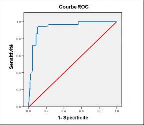
Performance diagnostique de l’interféron gamma dans l’identification de l’origine tuberculeuse des pleurésies exsudatives
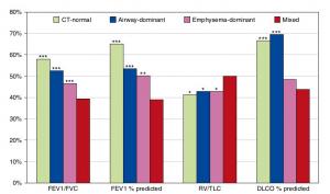
A Mixed Phenotype of Airway Wall Thickening and Emphysema Is Associated with Dyspnea and Hospitalization for Chronic Obstructive Pulmonary Disease.

Radiological Approach to Asthma and COPD-The Role of Computed Tomography.

Significant annual cost savings found with UrgoStart in UK and Germany
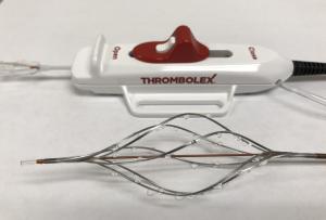
Thrombolex announces 510(k) clearance of Bashir catheter systems for thromboembolic disorders
Phone: (028) 3981 2678
Mobile: 0903 839 878 - 0909 384 389
