I.Anesthesia
Tumescent anesthetic, when used in phlebology, describes the use of large volumes of dilute anesthetic solutions that are infiltrated into the perivenous space of the veins to be treated.
The rationale behind the use of large volume tumescent anesthesia for ELA include its use as a local anesthetic, its ability to empty the vein to maximize the contact of the thermal device and the vein wall for efficient thermal transfer to the vein wall, and providing a protective heat sink around the treated vein to minimize heating of adjacent structures.
ELA is usually performed with a dilute tumescent anesthetic solution of lidocaine with or without epinephrine in normal saline, often buffered with sodium bicarbonate (a concentration of 0.1% lidocaine is typically used with an average volume of about 5–10 mL/cm of treated vein). This should be delivered with ultrasound guidance into the perivenous space (saphenous sheath) of the vein to be treated. It can be injected either manually or with an infusion pump, such that upon completion of the process the vein is surrounded along its entire treated length with the anesthetic fluid, as demonstrated in the image below.
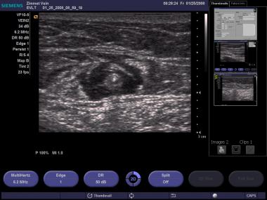
Transverse ultrasound image of tumescent anesthetic fluid surrounding centrally located great saphenous vein and laser fiber/sheath.
Although the maximum safe dosage of lidocaine using the tumescent technique for venous procedures is not well studied, 35 mg/kg with epinephrine has been reported as safe in the plastic surgical literature. However, the US Food and Drug Administration (FDA)–reviewed circulars accompanying units of lidocaine state a maximum dose of 5 mg/kg without and 7 mg/kg with epinephrine with each use. Toxicity may occur related to the dose of lidocaine and or epinephrine. Care should be used in patients who are likely to be more sensitive to the dose of these drugs, including elderly persons. When using epinephrine, the use of ECG monitoring may be prudent.
II.Equipment
Basic equipment and supplies for ELA are listed below:
- Procedure table that can tilt to Trendelenburg and reverse Trendelenburg
- DUS with at least a 7.5 MHz transducer
- Sterile gowns, gloves, masks, drapes, gauze
- Ultrasound gel, sterile ultrasound probe and cord cover
- Antiseptic preparation fluid
- Local anesthetic
- No. 11 scalpel blade, 18-gauge needle, 15º ophthalmic blade, or punch biopsy device
- 18- to 21-gauge needle for percutaneous entry
- 21- to 25-gauge needle for administration of tumescent anesthesia
- Syringes
- Normal saline
- Compression stockings
A foot pedal controlled tumescent anesthetic injection pump can be used to infuse the perisaphenous anesthetic as an alternative to hand injection. Venous access kits that allow the use of a less traumatic 21-gauge needle to insert a 0.018-in guidewire are useful when accessing small veins but do add expense to the procedure. These kits include a 4 or 5F sheath with a dilator tapered to the 0.018-in guidewire. After the catheter and dilator are inserted, the dilator and 0.018-in guidewire can be removed to allow the placement of a standard 0.035-in guidewire. These micropuncture kits are marketed by a variety of vendors.
Additional materials required to perform ELA include the laser generator and sterile
laser fiber (see list below) and sheath long enough to cross the abnormal venous segment(s), usually included in a kit along with a guidewire. ELA is usually performed by placing a 4 or 5F sheath into the vein to be treated over a 0.035-in guidewire and then, after inserting a laser fiber into the sheath, withdrawing the sheath to expose the fiber tip. The sheaths are manufactured in multiple lengths and generally the sheath chosen is as long as or longer than the segment(s) to be treated. Sheaths that have a ruler imprinted on them make it easiest to monitor the rate at which they are withdrawn. In very straight veins, a laser fiber can be advanced beyond its sheath to the starting point of ablation. Kits are now available with blunt-tip laser fibers to facilitate this. However, advancement through the sheath is recommended in tortuous veins to avoid passing the fiber through the vein wall.
III.ELA tools
ELA can be performed using any of the following wavelengths. Generators and laser fiber kits for use are marketed by multiple vendors, as follows:
Endovenous laser wavelengths commercially available include:
- 810 nm (AngioDynamics Queensbury, NY)
- 940 nm (Dornier MedTech Americas, Inc, Kennesaw, Ga)
- 980 nm (Biolitec, Inc, East Longmeadow, Mass)
- 1064 nm (Sharplan, Inc., NJ)
- 1320 nm (CoolTouch, Roseville, Calif)
- 1470 nm (Biolitec, Angiodynamics)
Laser ablation has been primarily performed with 810-µm bare-tipped fibers, which are premarked to allow the operator to know when the fiber is tip-to-tip with the end of the sheath and when the laser extends a fixed distance beyond the sheath tip. Although many of the original fibers were bare-tipped, many of the currently used fibers are jacketed with ceramic or metal, which, in theory, may decrease vein wall perforation and increase the effective diameter of the fiber, resulting in a decrease in the power density and changing the fiber from a cutting mode into a coagulation mode.Anecdotally, with similar power settings and pull-back rates, there is less pain and bruising, although the long-term success has not been characterized to see if this results in a trade off in efficacy. Limited data are available that compare the different configurations, but anecdotally it is thought that higher, water-specific wavelengths produce less postprocedure pain with equivalent outcomes.
IV.Positioning
Access to the target vein should be performed with the patient in the supine position. The use of a reverse Trendelenburg position (feet down) in order to increase pressure in the target vein and increase the likelihood of a successful puncture is advisable, especially with small-diameter veins. Once the sheath and laser fiber are inserted as described below, the patient is positioned flat and then in the Trendelenburg position after positioning the laser fiber at the desired starting location. The Trendelenburg position helps to empty the vein and improve energy transfer from the fiber to the vein wall. This is particularly important at the upper end of the greater saphenous vein (GSV), where the vein diameter is larger and the vein is less susceptible to spasm.
Source emedicine.com
DUC TIN CARDIO SURGICAL CLINIC
Tin tức liên quan
Performance diagnostique de l’interféron gamma dans l’identification de l’origine tuberculeuse des pleurésies exsudatives
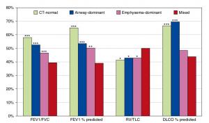
A Mixed Phenotype of Airway Wall Thickening and Emphysema Is Associated with Dyspnea and Hospitalization for Chronic Obstructive Pulmonary Disease.

Radiological Approach to Asthma and COPD-The Role of Computed Tomography.
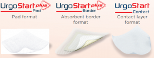
Significant annual cost savings found with UrgoStart in UK and Germany
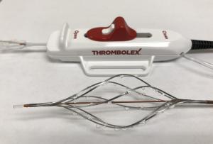
Thrombolex announces 510(k) clearance of Bashir catheter systems for thromboembolic disorders
Phone: (028) 3981 2678
Mobile: 0903 839 878 - 0909 384 389
