Complications
Adverse events and complications
Adverse events following ELA occur, but almost all are minor. Ecchymosis over the treated segment frequently occurs and normally lasts for 7-14 days. About one week after ELA, the treated vein may develop a feeling of tightness similar to that after a strained muscle.
This transient discomfort, likely related to inflammation in the treated vein segment, is self-limited and may be ameliorated with the use of nonsteroidal anti-inflammatory drugs (NSAIDs), ambulation, stretching, and graduated compression stockings. Both of these side effects are more commonly described after ELA using existing laser protocols than for RFA, but the differences in severity are very small when studied objectively.
Superficial phlebitis is another uncommon side effect of ELA, being reported after about 5% of treatments as mentioned previously. There are no published reports of superficial phlebitis after ELA progressing to deep vein thrombosis and it has been managed in most series with NSAIDs, graduated compression stockings, and ambulation. Anecdotally, superficial phlebitis seems to be more common in larger diameter tributary varicose veins or in varicose veins that have their inflow and outflow ablated by ELA. Concurrent phlebectomy of these veins at the time of ELA has been recommended to decrease the risk of this side effect, but at this point no data substantiate this claim.
More significant adverse events reported following ELA include neurologic injuries, skin burns, and DVT. The overall rate of these complications has been shown to be higher in low-volume centers than high-volume centers. The nerves at highest risk include the saphenous nerve, adjacent to the GSV below the mid-calf perforating vein, and the sural nerve adjacent to the SSV in the mid and lower calf. Both of these nerves have only sensory components. The most common manifestation of a nerve injury is a paresthesia or dysesthesia, most of which is transient. The nerve injuries can occur with the trauma associated with catheter introduction, during the delivery of tumescent anesthesia, or by thermal injury related to heating of the perivenous tissues.
Tumescent anesthesia has been demonstrated to reduce perivenous temperatures with laser and RF ablation. The delivery of the perivenous fluid is felt to be responsible for the low rate of cutaneous and neurologic thermal injuries seen in the series of patients treated using perivenous fluid. Neurologic injuries are seen after truncal vein removal and are related to injury to nerves adjacent to the treated vein. The incidence of these adverse events are related to the degree to which objective testing is performed to identify them. In general, paresthesias caused by ELA are usually temporary with the rate of permanent paresthesias typically reported for GSV and SSV as 0–10%.
The one-week paresthesia rate following RFA was shown to decrease from 15% to 9% after the introduction of tumescent anesthesia. Patients treated with laser ELA performed without tumescent anesthetic infiltrations also demonstrated a high rate of such injuries. Evidence suggests a higher rate of nerve injuries when treating the below knee GSV as compared with the above knee segment and the SSV. Treatment of the below knee GSV or lower part of the SSV may be necessary in many patients to treat to eliminate symptoms or skin disease caused by reflux to the ankle.
A retrospective review demonstrated that below knee laser ablation can be performed with an 8% rate of mild but permanent paresthesias with adequate amounts of tumescent anesthesia. This data also suggests that sparing the treatment of the distal 5–10 cm may have clinical benefit and reduce saphenous nerve injury risk in patients with reflux to the medial malleolus. Skin burns following ELA have been reported. Skin burns are fortunately relatively rare and seem to be avoidable with adequate tumescent anesthesia. The rate of skin burn in 1 series using RFA was 1.7% before and 0.5% after the initiation of the use of tumescent technique during RFA. The early experience had rates as high as 4% that decreased to almost 0% as the use of tumescent anesthesia became a standard of practice.
DVT following ELA is unusual. DVT can occur as an extension of thrombus from the treated truncal vein across the junctional connection into the femoral or popliteal veins. The reported rates of junctional thrombosis following GSV ELA varies widely. This variability may relate to the time of the follow-up examination and the methods used. Most published series using early DUS (around 72 hours or less after ELA) document a proximal extension for the GSV around 1%.
The risk of venous thromboembolism (VTE) is higher in patients with a history of prior DVT or phlebitis, CEAP (clinical, etiological, anatomical and pathological) classification of 3 or greater, and male sex.Endothermal heat-induced thrombosis (EHIT) defines the extent of superficial thrombosis and its extension into the deep venous system as proposed by Kabnick.EHIT 1 represents thrombus up to the SFJ, EHIT 2 represents thrombus extending into the femoral vein occupying less than 50% of luminal diameter, EHIT 3 represents thrombus occupying greater than 50% of femoral vein luminal diameter, and EHIT 4 represents occlusive thrombus in the femoral vein. EHIT 1 is treated conservatively. If identified, EHIT 2 is usually treated with anticoagulation (full or prophylactic intensity are both used), although some advocate early re-examination and conservative care for more minor forms. EHIT 3 and 4, which are much less common, probably merit full anticoagulation.
Those performing the DUS at a later interval identify a lower rate of EHIT. Possibly, the rates are different for different operators with different protocols or the proximal extension of thrombus may be self-limited and may resolve by 1 month without a clinical event. Pooling data from several sources suggest that the incidence is approximately 0.3% after ELA. This type of DVT is almost universally asymptomatic. The significance of this type of thrombus extension into the femoral vein seems to be different from that found with native GSV thrombosis with extension or when compared with typical femoral vein thrombosis.
The incidence of junctional extension of thrombus after SSV ablation has also been described to be low (0-6%). In one study, the rate of popliteal extension of SSV thrombus at 2-4 days after ELA was related to the anatomy of the SPJ. The incidence at 48-72 hours of follow-up was 0% when no SPJ existed, 3% when a thigh extension exists, but 11% when no thigh extension could be identified just above the SPJ. Heparin was used to treat identified thrombus extensions and all regressed. No published data are available on conservative management of transjunctional thrombus extension at either the SPJ or SFJ. However, given that popliteal or femoral vein obstruction develops in significantly less than 1% of patients, including in those series where DUS is not done until 1 month after ELA, the practice of performing early DUS surveillance and aggressive anticoagulation of such findings is controversial.
Neovascularity at the SFJ after ELA, as a form of recurrence of varicose veins, seems to be rare at 1- to 3-year follow-up. Neovascularization was seen in only 2 of the 1222 limbs followed for up to 5 years in an industry-sponsored registry of patients treated with RFA. Longer follow-up may be necessary to feel confident with this observation. However, neovascularization is common and often an early event following high-ligation and stripping (HL/S). Neovascularization may be less common following endovenous procedures because the junctional tributary flow, which was usually ligated at their confluence with the SFJ, is generally not affected with GSV ELA.
Anecdotal reports of laser fiber fracture or retained venous access sheaths have been made to the device manufacturers and a case report exists describing a retained vascular sheath after laser ablation. Respecting the fragile glass laser fibers and being gentle with its handling should help minimize laser fiber fractures. The possibility of a laser fiber fracture should be considered with the removal of the device in each case. Care to deliver thermal energy only beyond the introducer sheath and away from any other parallel placed sheaths when treating 2 veins during the same procedure is essential to avoid severing segments of these catheters. No specific management recommendations of retained intravenous laser fiber or sheath fragments can be made based on the data. However, anecdotally, retained short segments of the distal end of the laser fiber seem to be well tolerated without incident and efforts to remove them may be more prone to adverse events than managing them conservatively.
A case report of an arteriovenous fistula (AVF) between a small popliteal artery branch near the SPJ and the SSV exists. Anecdotal references have been made of additional AVFs between the proximal GSV and the contiguous superficial external pudendal artery. Although thought to be related to a heat-induced injury caused by the thermal device, an AVF could be caused by a needle injury during tumescent anesthetic administration. Ways to minimize the risk of these AVFs include careful advancement of the intravascular devices, atraumatic delivery of the tumescent anesthetic, the use of copious amounts of tumescent fluid, and avoidance of treating the subfascial portion of the SSV where popliteal artery branches exist adjacent to the SSV.
Source emedicine.com
DUC TIN SURGICAL CLINIC
Tin tức liên quan
Performance diagnostique de l’interféron gamma dans l’identification de l’origine tuberculeuse des pleurésies exsudatives
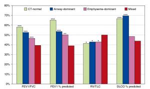
A Mixed Phenotype of Airway Wall Thickening and Emphysema Is Associated with Dyspnea and Hospitalization for Chronic Obstructive Pulmonary Disease.
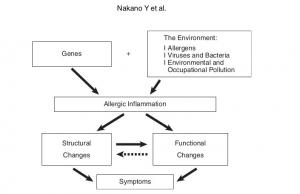
Radiological Approach to Asthma and COPD-The Role of Computed Tomography.
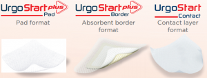
Significant annual cost savings found with UrgoStart in UK and Germany
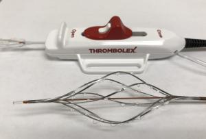
Thrombolex announces 510(k) clearance of Bashir catheter systems for thromboembolic disorders
Phone: (028) 3981 2678
Mobile: 0903 839 878 - 0909 384 389
