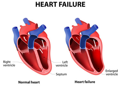
Your doctor will diagnose heart failure based on your medical and family histories, a physical exam, and test results. The signs and symptoms of heart failure also are common in other conditions. Thus, your doctor will:
- Find out whether you have a disease or condition that can cause heart failure, such as coronary heart disease (CHD), high blood pressure, or diabetes
- Rule out other causes of your symptoms
- Find any damage to your heart and check how well your heart pumps blood
Early diagnosis and treatment can help people who have heart failure live longer, more active lives.
Medical and Family Histories
Your doctor will ask whether you or others in your family have or have had a disease or condition that can cause heart failure.
Your doctor also will ask about your symptoms. He or she will want to know which symptoms you have, when they occur, how long you've had them, and how severe they are. Your answers will help show whether and how much your symptoms limit your daily routine.
Physical Exam
During the physical exam, your doctor will:
- Listen to your heart for sounds that aren't normal
- Listen to your lungs for the sounds of extra fluid buildup
- Look for swelling in your ankles, feet, legs, abdomen, and the veins in your neck
Diagnostic Tests
No single test can diagnose heart failure. If you have signs and symptoms of heart failure, your doctor may recommend one or more tests.
Your doctor also may refer you to a cardiologist. A cardiologist is a doctor who specializes in diagnosing and treating heart diseases and conditions.
EKG (Electrocardiogram)
An EKG is a simple, painless test that detects and records the heart's electrical activity. The test shows how fast your heart is beating and its rhythm (steady or irregular). An EKG also records the strength and timing of electrical signals as they pass through your heart.
An EKG may show whether the walls in your heart's pumping chambers are thicker than normal. Thicker walls can make it harder for your heart to pump blood. An EKG also can show signs of a previous or current heart attack.
Chest X Ray
A chest x ray takes pictures of the structures inside your chest, such as your heart, lungs, and blood vessels. This test can show whether your heart is enlarged, you have fluid in your lungs, or you have lung disease.
BNP Blood Test
This test checks the level of a hormone in your blood called BNP. The level of this hormone rises during heart failure.
Echocardiography
Echocardiography (echo) uses sound waves to create a moving picture of your heart. The test shows the size and shape of your heart and how well your heart chambers and valves work.
Echo also can identify areas of poor blood flow to the heart, areas of heart muscle that aren't contracting normally, and heart muscle damage caused by lack of blood flow.
Echo might be done before and after a stress test (see below). A stress echo can show how well blood is flowing through your heart. The test also can show how well your heart pumps blood when it beats.
Doppler Ultrasound
A Doppler ultrasound uses sound waves to measure the speed and direction of blood flow. This test often is done with echo to give a more complete picture of blood flow to the heart and lungs.
Doctors often use Doppler ultrasound to help diagnose right-side heart failure.
Holter Monitor
A Holter monitor records your heart's electrical activity for a full 24- or 48-hour period, while you go about your normal daily routine.
You wear small patches called electrodes on your chest. Wires connect the patches to a small, portable recorder. The recorder can be clipped to a belt, kept in a pocket, or hung around your neck.
Nuclear Heart Scan
A nuclear heart scan shows how well blood is flowing through your heart and how much blood is reaching your heart muscle.
During a nuclear heart scan, a safe, radioactive substance called a tracer is injected into your bloodstream through a vein. The tracer travels to your heart and releases energy. Special cameras outside of your body detect the energy and use it to create pictures of your heart.
A nuclear heart scan can show where the heart muscle is healthy and where it's damaged.
A positron emission tomography (PET) scan is a type of nuclear heart scan. It shows the level of chemical activity in areas of your heart. This test can help your doctor see whether enough blood is flowing to these areas. A PET scan can show blood flow problems that other tests might not detect.
Cardiac Catheterization
During cardiac catheterization (KATH-eh-ter-ih-ZA-shun), a long, thin, flexible tube called a catheter is put into a blood vessel in your arm, groin (upper thigh), or neck and threaded to your heart. This allows your doctor to look inside your coronary (heart) arteries.
During this procedure, your doctor can check the pressure and blood flow in your heart chambers, collect blood samples, and use x rays to look at your coronary arteries.
Coronary Angiography
Coronary angiography (an-jee-OG-rah-fee) usually is done with cardiac catheterization. A dye that can be seen on x ray is injected into your bloodstream through the tip of the catheter.
The dye allows your doctor to see the flow of blood to your heart muscle. Angiography also shows how well your heart is pumping.
Stress Test
Some heart problems are easier to diagnose when your heart is working hard and beating fast. During stress testing, you exercise to make your heart work hard and beat fast.
You may walk or run on a treadmill or pedal a bicycle. If you can't exercise, you may be given medicine to raise your heart rate.
Heart tests, such as nuclear heart scanning and echo, often are done during stress testing.
Cardiac MRI
Cardiac MRI (magnetic resonance imaging) uses radio waves, magnets, and a computer to create pictures of your heart as it's beating. The test produces both still and moving pictures of your heart and major blood vessels.
A cardiac MRI can show whether parts of your heart are damaged. Doctors also have used MRI in research studies to find early signs of heart failure, even before symptoms appear.
Thyroid Function Tests
Thyroid function tests show how well your thyroid gland is working. These tests include blood tests, imaging tests, and tests to stimulate the thyroid. Having too much or too little thyroid hormone in the blood can lead to heart failure.
Source emedicine.com
DUC TIN SURGICAL CLINIC
Tin tức liên quan
Performance diagnostique de l’interféron gamma dans l’identification de l’origine tuberculeuse des pleurésies exsudatives
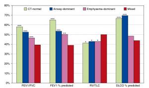
A Mixed Phenotype of Airway Wall Thickening and Emphysema Is Associated with Dyspnea and Hospitalization for Chronic Obstructive Pulmonary Disease.

Radiological Approach to Asthma and COPD-The Role of Computed Tomography.

Significant annual cost savings found with UrgoStart in UK and Germany
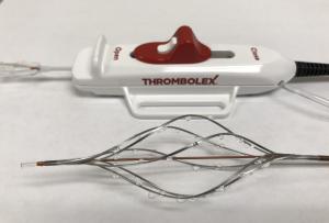
Thrombolex announces 510(k) clearance of Bashir catheter systems for thromboembolic disorders
Phone: (028) 3981 2678
Mobile: 0903 839 878 - 0909 384 389
