


The most optimal gene delivery system for cardiac therapeutic applications in humans will combine a clinically advantageous physical delivery route with the rAAV serotype that has the highest natural or engineered affinity for the targeted cardiac cell type.
Additional use of a cell-type biased expression cassette (see Cardiac-Targeted and Regulated Expression Systems section for details) is expected to confine therapeutic gene expression to the targeted organ. Ideally, this procedure is safe and efficient when it comes to translation in humans. In the following section, we will briefly discuss delivery methods in small animals and focus on administration routes in preclinical large animal readily extrapolatable to humans or already used in phase I/II trials.
Myocardial Gene Delivery in Small Animal Models
Intravenous gene delivery via tail, jugular, or retro-orbital veins is the least invasive technique to administer a vector for myocardial gene delivery, but highly dependent on affinity of the vector toward cardiomyocytes. A recent study has shown that intravenous application of 1×10¹¹ viral particles of rAAV9 in wild-type C57B/6 mice harboring the β-adrenergic receptor kinase C terminus (βARKct) cDNA driven by a cardiac-biased promoter resulted in homogeneous cardiac expression confined to cardiomyocytes. This approach is deemed highly useful for proof-of-concept but also for dose-dependency studies of DNA and RNA therapeutics in rodent models yielding necessary PK/PD data for definition of therapeutic window and threshold for adverse effects (see PK and PD of Cardiac Gene Therapeutics section for details). Although another study has reported tropism of AAV9 toward cardiac and skeletal muscle in nonhuman primates,improved transductional targeting is needed until this technique can be considered for use in human-relevant large animal HF models.
Intracoronary gene delivery in small animals has been used widely to enhance viral transduction efficiency in hearts via cardiac-directed application. The ascending aorta is cross-clamped, and injection of the vector solution into the aortic root or into the left ventricle via catheter enables antegrade intracoronary infusion. Reported myocardial transduction rate, assessed by reporter genes, varied between 20% and 50% when 1×10¹⁰ to 1×10¹² viral particles both of rAAV2, rAAV6, and rAd5 were used. Efficiency can further be increased by infusion of histamine,adenosine, and various other NO donors to enhance endothelial permeability.
Pericardial gene delivery has also been tested for rAd in small animal models and offers the advantage of targeted gene delivery to pericardial cells. The epicardial layer limits transduction of adjacent myocardium, and use of various pharmacological agents and proteolytic enzymes to overcome the barrier bears potential for cardiac damage. The technique thus yields insufficient cardiomyocyte transduction for proof-of-concept studies.
Myocardial Gene Delivery in Translational Studies and Large Animal Models
Antegrade coronary artery gene delivery is a minimally invasive percutaneous catheter-based injection into the coronary arteries (Figure 4A). The technique is clinical routine and, principally, gene delivery can be safely directed to each area of the heart. However, endothelial dysfunction, atherosclerosis, and occlusion of coronary arteries in humans with coronary artery disease might limit myocardial transduction efficiency at the cost of greater viral spill-over. Passage time of the delivered vector further depending on regional perfusion differences is usually short. A significant amount of the applied vector dosage is usually washed out into systemic circulation where it is subject to renal excretion or hepatic transduction. A first approach to enhance myocardial retention is temporary occlusion of the coronary artery and the corresponding coronary vein (Figure 4Band 4C), which increased myocardial gene expression significantly, but bears potential for cardiac damage. Some studies showed only a few β-gal-positive–stained cardiomyocytes after intracoronary Adβ-gal delivery in pigs at a dosage of 5×10¹² viral particles. Others reported on increased left ventricular (LV) function in pig models of congestive HF when AAV1-SERCA2a or Ad5-ACVI was delivered via coronary arteries at a dosage of 1×10¹³ and 1.4×10¹² viral particles without reported myocardial transduction rates

Figure 4.
Clinically applicable catheter-based cardiac-targeted gene delivery modes. Antegrade intracoronary perfusion (A) is currently used in phase I and phase II clinical heart failure gene therapy trials. Other forms (B–E) have successfully used in preclinical studies being either clinical routine or readily clinically applicable. Adapted with permission from Tilemann et al.21 Authorization for this adaptation has been obtained both from the owner of the copyright in the original work and from the owner of copyright in the translation or adaptation.
Various chemical agents have been tested to increase myocardial transduction, eventually advocating NO-dependent enhancement of vascular permeability and virus transmigration. Periprocedural intracoronary NO donor delivery, such as nitro-glycerine, was routinely used in the CUPID and Ad5.hAC6 Gene Transfer for Congestive Heart Failure trials. In the quest for ideal myocardial transduction, a recirculating, closed-loop system between the coronary sinus and the left main coronary artery using a peristaltic pump (V-Focus) has been developed (Figure 4D). Using V-Focus, AAV2-SERCA2a gene delivery in pacing-induced HF in adult sheep demonstrated dose-dependent (1×10¹⁰, 1×10¹², and 1×10¹³ DNAse-resistant particles) beneficial functional and structural effects, but myocardial transduction rates were not available. The ambitious procedure might show its advantage, optimized vector dosage with efficient myocardial transduction, when handling can be further simplified and costs diminished to meet demands of a daily clinical routine. Because of superior safety and feasibility, clinical HF gene therapy trials testing SERCA2a and AC6 use percutaneous catheter-based antegrade vector injection into the coronary arteries. Naturally, evidence of myocardial transduction cannot be provided but results of phase IIa of the CUPID trial are encouraging. However, efficiency in patients with coronary artery disease is a matter of ongoing debate, and anticipated results of phase III of the CUPID trial might provide clarification.
Retrograde coronary venous gene delivery is based on percutaneous minimally invasive retrograde myocardial gene delivery via the coronary venous system (Figure 4E). The clinical use of this method is increasing because of the demand for cardiac resynchronization therapy. The anterior cardiac vein is sealed by a balloon, whereas the vector solution is retroinfused via catheter so that hibernating, viable myocardium can be targeted when corresponding coronary arteries are occluded. Passage time is significantly prolonged as compared with antegrade injection, and myocardial gene delivery was further enhanced when coronary arterial blood flow was temporarily blocked. When used in human-relevant large animal HF models, retrograde coronary venous delivery of 1.5×10¹³AAV9-S100A1 and 1.0×10¹³ AAV6-βARKct particles was safe, rescued cardiac dysfunction, and reversed remodeling because of sufficient myocardial transduction. Interestingly, significant myocardial transfection was also achieved by retrograde delivery of plasmid DNA, which deserves further consideration.
Percutaneous intramyocardial injection describes a nonsurgical minimally invasive technique of intramyocardial gene delivery that requires a catheter with a retractable injection needle and imaging guidance modalities for determining the injection site. Among mapping systems, fluoroscopy, echocardiography, and MRI, the NOGA electromechanical mapping system is the most commonly used guiding system used in clinical angiogenesis trials. With therapeutic DNA/RNA formulations acting inside cardiomyocytes, the system might not allow sufficient transduction efficiency but might act favorably when a therapeutic paracrine factor can be used.
Surgical administration of vectors is convenient in patients undergoing open-heart surgery. Antegrade or retrograde gene delivery via coronary arteries during aortic cross-clamping for 30 minutes yielded highly efficient and selective myocardial gene expression. Refinement of selective retrograde coronary sinus infusion and recirculation of an AAV6-βARKct vector resulted in robust, although not quantified, gene expression and increased adrenergic reserve in sheep. In addition, the exposed heart is amenable to targeted intramyocardial injection of therapeutic vectors.
Source ahajournals
DUC TIN SURGICAL CLINIC
Tin tức liên quan
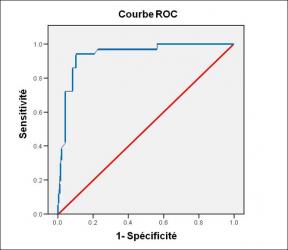
Performance diagnostique de l’interféron gamma dans l’identification de l’origine tuberculeuse des pleurésies exsudatives
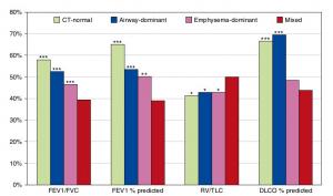
A Mixed Phenotype of Airway Wall Thickening and Emphysema Is Associated with Dyspnea and Hospitalization for Chronic Obstructive Pulmonary Disease.

Radiological Approach to Asthma and COPD-The Role of Computed Tomography.
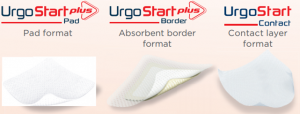
Significant annual cost savings found with UrgoStart in UK and Germany
Thrombolex announces 510(k) clearance of Bashir catheter systems for thromboembolic disorders
Phone: (028) 3981 2678
Mobile: 0903 839 878 - 0909 384 389