Abdominal aortic aneurysms (AAAs) are relatively common and are potentially life-threatening. Aneurysms are defined as a focal dilatation in an artery, with at least a 50% increase over the vessel’s normal diameter. Thus, enlargement of the diameter of the abdominal aorta to 3 cm or more fits the definition.
AAA usually results from degeneration in the media of the arterial wall, leading to a slow and continuous dilatation of the lumen of the vessel. Uncommon causes include infection, cystic medial necrosis, arteritis, trauma, inherited connective-tissue disorders, and anastomotic disruption.
AAAs generally affect elderly white men. Smoking appears to be the risk factor most strongly associated with AAA. In addition to increasing age and male sex, other factors include increased height, weight, body mass index, and body surface area. A familiar clustering has been noted in 15-25% of patients undergoing surgical repair of AAA. Female sex, African American race, and the presence of diabetes mellitus are negatively associated with AAA.
Most AAAs are asymptomatic, and many are detected as an incidental finding on diagnostic imaging obtained for other reasons. There is a wide spectrum of clinical presentations, and AAA should be considered in the differential diagnosis for a number of symptoms.
Ultrasonography is the standard imaging tool for AAA. Bedside emergency ultrasonography should be performed immediately if AAA is suspected.
Treatment of abdominal aortic aneurysms (AAAs) is with surgical repair. When indicated, unruptured aneurysms can undergo elective repair (see Treatment). The combination of ultrasonographic screening, reduced preoperative risk, and new minimally invasive techniques extend aortic aneurysm treatment into an increasingly elderly population. If an AAA ruptures, emergency surgical repair is required.
Anatomy
The abdominal aorta has three distinct tissue layers: intima, media, and adventitia. The intima is composed of the classic endothelial layer. The media comprises smooth muscle cells surrounded by elastin, collagen, and proteoglycans; to a great extent, this layer is responsible for the structural and elastic properties of the artery. The adventitia consists primarily of collagen but also contains a variety of cells (including fibroblasts and immunomodulatory cells), as well as adrenergic nerves.
The diameter of the aorta decreases from its thoracic portion to its abdominal and infrarenal portions. A normal aorta shows a reduction in medial elastin layers from the thoracic portion to the abdominal portion. Elastin content and collagen content are also reduced.
Most AAAs begin below the renal arteries and end above the iliac arteries. The size, shape, and extent of AAAs vary considerably. Like aneurysms of the thoracic aorta, AAAs may be broadly described as either fusiform (circumferential) or saccular (more localized). However, these descriptions represent two points on a continuum, and lesions that fall between the two points exist.
The important surgical and endovascular anatomic considerations include associated renal and visceral artery involvement (either occlusive disease or involved in the aneurysm process) and the iliac artery (either occlusive disease or aneurysms). The length of the infrarenal aortic neck is important in helping determine the surgical approach (ie, retroperitoneal or transabdominal) and the location of the aortic cross-clamp.
Consideration of hypogastric artery (internal iliac) outflow is important in planning surgical repair. Loss of blood flow from the hypogastric artery may result in impotence in males and sigmoid colon ischemia with necrosis.
Inflammatory aneurysms represent a subsegment of AAA and are characterized by a thick inflammatory peel. These aneurysms are associated with retroperitoneal fibrosis and adhesion of the duodenum and fibrosis (see the image below).
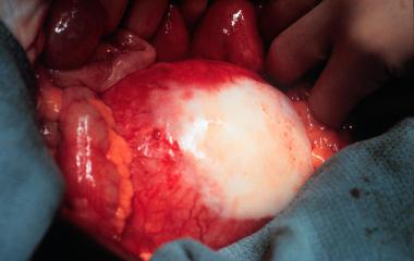
Aneurysm with retroperitoneal fibrosis and adhesion of duodenum.
Source emedicine.com
Duc Tin Surgical Clinic
Tin tức liên quan
Performance diagnostique de l’interféron gamma dans l’identification de l’origine tuberculeuse des pleurésies exsudatives
A Mixed Phenotype of Airway Wall Thickening and Emphysema Is Associated with Dyspnea and Hospitalization for Chronic Obstructive Pulmonary Disease.
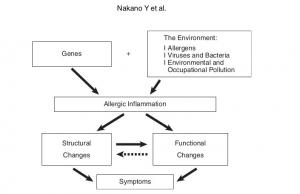
Radiological Approach to Asthma and COPD-The Role of Computed Tomography.
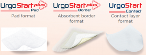
Significant annual cost savings found with UrgoStart in UK and Germany
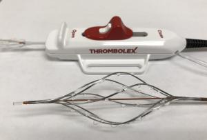
Thrombolex announces 510(k) clearance of Bashir catheter systems for thromboembolic disorders
Phone: (028) 3981 2678
Mobile: 0903 839 878 - 0909 384 389
