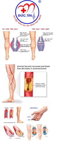
Most clinically significant AAAs are palpable upon routine physical examination; however, the sensitivity of palpation depends on the experience of the examiner, the size of the aneurysm, and the size of the patient.
In one study, 38% of AAA cases were detected on the basis of physical examination findings, whereas 62% were detected incidentally on radiologic studies obtained for other reasons.
Abdominal examination includes palpation of the aorta and estimation of the size of the aneurysm. AAAs are palpated in the upper abdomen; the aorta bifurcates into the iliac arteries just above the umbilicus. The clinician need not be afraid of properly palpating the abdomen, because there is no evidence to indicate that aortic rupture can be precipitated by this maneuver.
Whereas the abrupt onset of pain due to rupture of an AAA may be quite dramatic, the associated physical findings may be very subtle. Patients may have normal vital signs in the presence of a ruptured AAA as a consequence of retroperitoneal containment of hematoma.
The presence of a pulsatile abdominal mass (see the image below) is virtually diagnostic of an AAA but is found in fewer than 50% of cases. It is more likely to be noted with a ruptured aneurysm. In an obese abdomen, an AAA is more difficult to palpate. Even in patients known to have an aneurysm, vascular surgeons are unable to palpate a pulsatile mass while preparing the patient for surgery in 25% of cases.

Pulsatile abdominal mass.
Occasionally, an overlying mass (pancreas or stomach) may be mistaken for an AAA. An abdominal bruit is nonspecific for an unruptured aneurysm, but the presence of an abdominal bruit or the lateral propagation of the aortic pulse wave can offer subtle clues and may be more frequently found than a pulsatile mass. Bruits may also indicate the presence of renal or visceral artery stenosis; a thrill is possible with aortocaval fistulae. Patients with popliteal artery aneurysms frequently have AAAs (25-50% of cases).
Misdiagnosis is fairly common because the classic presentation of pain associated with hypotension, tachycardia, and a pulsatile abdominal mass is present in less than 30-50% of cases. The leading misdiagnosis is renal colic; dissection of the renal artery may produce flank pain and hematuria.
Normally, systolic blood pressures are higher in the thigh than in the arm. In patients with AAA, this relation may be reversed. Bilateral upper-extremity blood pressures should be measured in patients with AAAs. Upper-extremity blood pressures that differ from each other by more than 30 mm Hg indicate subclavian artery stenosis, and perioperative monitoring is important. Cervical bruits may indicate carotid artery stenosis. Hypertension may trigger a workup for renal artery stenosis.
Femoral/popliteal pulses and pedal (dorsalis pedis or posterior tibial) pulses should be palpated to determine if an associated aneurysm (femoral/popliteal) or occlusive disease exists. Flank ecchymosis (Grey Turner sign) represents retroperitoneal hemorrhage.
Source emedicine.com
Duc Tin Surgical Clinic
Tin tức liên quan
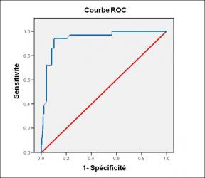
Performance diagnostique de l’interféron gamma dans l’identification de l’origine tuberculeuse des pleurésies exsudatives
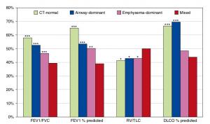
A Mixed Phenotype of Airway Wall Thickening and Emphysema Is Associated with Dyspnea and Hospitalization for Chronic Obstructive Pulmonary Disease.

Radiological Approach to Asthma and COPD-The Role of Computed Tomography.

Significant annual cost savings found with UrgoStart in UK and Germany
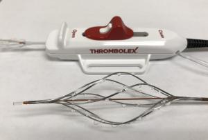
Thrombolex announces 510(k) clearance of Bashir catheter systems for thromboembolic disorders
Phone: (028) 3981 2678
Mobile: 0903 839 878 - 0909 384 389
