CT scanning accurately demonstrates dilation of the aorta and involvement of major branch vessels proximally and distally. This information helps in determining the appropriate intervention, which may be either surgical or endovascular repair. (See the image below.)
CT demonstrates an abdominal aortic aneurysm. The aneurysm was noted during workup for back pain, and CT was ordered after the abdominal aortic aneurysm was identified on radiographs. No evidence of rupture is seen.
CT also shows the other organs in the abdomen and demonstrates involvement or displacement of organs that can confuse the clinical picture. The location and number of the renal arteries, caliber of the aneurysm, degree of calcification, lengths of the neck and iliac artery, 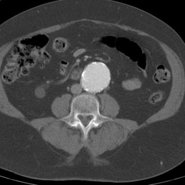 and presence of mural thrombus are readily assessed. CTA allows multiplanar assessment of the aneurysm and associated relevant vessels (visceral arteries, iliac and femoral arteries).
Degree of confidence
CT has emerged as the diagnostic imaging standard for the evaluation of AAA, with an accuracy that approaches 100%. A well-performed CT examination can reveal the extent of the aneurysm, as well as the involvement of other organs. Intravenously administered contrast agent is needed to obtain the full benefit of CT; however, a nonenhanced study accurately depicts AAAs. Three-dimensional reconstructions of state-of-the-art, multidetector-row, helical CT scans can help in preoperative planning and may replace the need for preoperative diagnostic angiography.
False positives/negatives
The administration of contrast material is essential for detecting dissection or ulceration of a vessel that might be missed without it. In the acute setting (eg, in a patient with back pain or an aneurysm), a false-positive diagnosis of rupture is possible if fluid resulting from another cause is seen in the abdomen. Conversely, an aneurysm or rupture can be missed in a patient who has recently undergone barium study, because artifact can obscure the aorta.
Source emedicine.com
Duc Tin Surgical Clinic
Tin tức liên quan
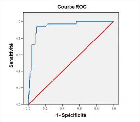
Performance diagnostique de l’interféron gamma dans l’identification de l’origine tuberculeuse des pleurésies exsudatives
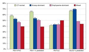
A Mixed Phenotype of Airway Wall Thickening and Emphysema Is Associated with Dyspnea and Hospitalization for Chronic Obstructive Pulmonary Disease.

Radiological Approach to Asthma and COPD-The Role of Computed Tomography.
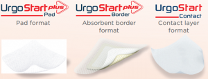
Significant annual cost savings found with UrgoStart in UK and Germany
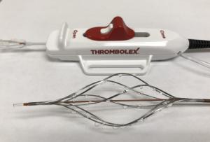
Thrombolex announces 510(k) clearance of Bashir catheter systems for thromboembolic disorders
Phone: (028) 3981 2678
Mobile: 0903 839 878 - 0909 384 389
