Overview
In 1840, Rayer first described the association between renal vein thrombosis (RVT) and nephrotic syndrome. Earlier reports, from postmortem examinations, had correctly cited infectious suppuration, malignancy, and trauma as likely causes of RVT.
Background
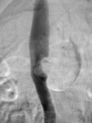
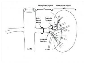
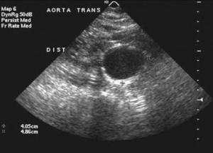
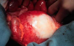
A renal artery aneurysm (RAA) is defined as a dilated segment of renal artery that exceeds twice the diameter of a normal renal artery. Symptomatic RAAs can cause hypertension, pain, hematuria, and renal infarction.
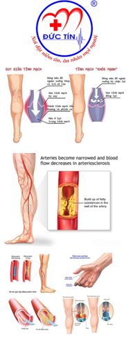
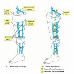
Varicose veins are simply dilated, tortuous veins of the subcutaneous/superficial venous system. However, the pathophysiology behind their formation is complicated and involves the concept of ambulatory venous hypertension.
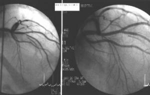
Coronary artery atherosclerosis is the single largest killer of men and women in the United States.
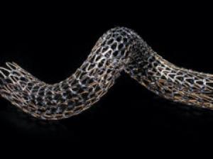
Gore has announced US Food and Drug Administration (FDA) approval of the Gore Tigris vascular stent, a dual-component stent with a unique fluoropolymer/nitinol design.
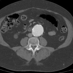
CT scanning accurately demonstrates dilation of the aorta and involvement of major branch vessels proximally and distally. This information helps in determining the appropriate intervention, which may be either surgical or endovascular repair. (See the image below.)
Radiography
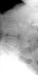
Calcification of the abdominal aortic wall is frequently evident on plain radiographs of the abdomen, as demonstrated in the images below.
Overview
In the United States, 15,000 deaths per year are attributed to abdominal aortic aneurysms (AAAs). Abdominal aortic aneurysms occur most commonly in individuals between 65 and 75 years of age.
Most clinically significant AAAs are palpable upon routine physical examination; however, the sensitivity of palpation depends on the experience of the examiner, the size of the aneurysm, and the size of the patient.
As noted (see Overview, Etiology), patients at greatest risk for abdominal aortic aneurysms (AAAs) are those who are older than 65 years and have peripheral atherosclerotic vascular disease.
AAA is thought to be a degenerative process of the aorta, the cause of which remains unclear. It is often attributed to atherosclerosis because these changes are observed in the aneurysm at the time of surgery.
AAAs arise as a result of a failure of the major structural proteins of the aorta (elastin and collagen). The inciting factors are not known, but a genetic predisposition clearly exists.
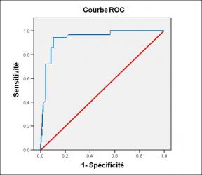
Performance diagnostique de l’interféron gamma dans l’identification de l’origine tuberculeuse des pleurésies exsudatives
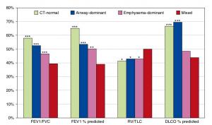
A Mixed Phenotype of Airway Wall Thickening and Emphysema Is Associated with Dyspnea and Hospitalization for Chronic Obstructive Pulmonary Disease.
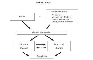
Radiological Approach to Asthma and COPD-The Role of Computed Tomography.

Significant annual cost savings found with UrgoStart in UK and Germany
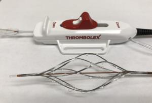
Thrombolex announces 510(k) clearance of Bashir catheter systems for thromboembolic disorders
Phone: (028) 3981 2678
Mobile: 0903 839 878 - 0909 384 389
